X
X
X
X
Back to Top

Case study: Diverse MPS symptoms and interdisciplinary approach lead to early diagnosis in 3-year-old with mucopolysaccharidosis VI (MPS VI)1

Age at diagnosis: 3 years

Time to diagnosis: <1 yeartrong>

Key diagnostic symptom: Heart murmur

Diagnosing doctor: Paediatric cardiology

Patient with MPS VI, ages 0 to 17

    Description1:

  • 3-year-old with short stature experienced recurring otitis media, joint issues and heart murmur; referred to paediatric cardiologist
  • Paediatric cardiologist recognised MPS VI and referred to geneticist
  • Diagnosed with MPS VI at 3 years of age
Early suspicion and a multidisciplinary approach can help drive timely MPS diagnosis and optimal long-term management.2

Summary:

Early diagnosis of MPS VI enabled early intervention and management, which are associated with improved clinical outcomes.2

  • Early clinical suspicion and a multidisciplinary approach are critical to diagnosis and long-term management2
  • In a 10-year follow-up resurvey study that included this patient, patients with MPS VI who received ERT had improved endurance and pulmonary function1,3
Early identification and initiation of ERT,a with accompanying active, multidisciplinary management, can dramatically improve long-term outcomes in MPS VI.2

Classic MPS symptoms – short stature, recurrent otitis media, skeletal and rheumatological manifestations – with accompanying cardiac involvement should prompt referral to a geneticist or metabolic centre.2

aIndividual outcomes in patients receiving ERT may vary.
Age 18 months:
  • Arrived at orphanage in Siberia, Russia
  • Adopted at age 3 and immigrated to the United States
Age 3:
  • Short stature noted by paediatrician; patient experienced frequent, recurrent otitis media and weakness in legs and was unable to sit straight or lift arms above head
  • Referred to paediatric cardiologist who recognised MPS VI and referred to geneticist for confirmatory testing and formal diagnosis
Age 4:
  • Multiple surgical interventions including shunt placement, hernial repair surgery, tonsillectomy/adenoidectomy
  • Occupational therapy and physical therapy initiated
Age 6:

Enzyme replacement therapy (ERT) initiated

Age 16:
  • Patient reported few rheumatological issues and can raise her arms above her head, sit with normal posture
  • Resolution of heart murmur
  • Infrequent ear, nose and throat involvement, with no reported airway obstruction or otitis media
  • No reported ocular or hearing issues
  • Significant improvement in quality of life: patient is pursuing professional singing career and rock climbing and aspiring to become a doctor
case-child1
case-child2
case-child3
case-child4
case-child5
case-child6

Case study: Postoperative paraplegia in a patient with Morquio A (MPS IVA) with spinal stenosis1

After an uneventful epidural anaesthesia in a patient with Morquio A and spinal stenosis, paraplegia became immediately apparent in the postanaesthesia care unit (PACU) setting1

    Description1:

  • 16-year-old patient with Morquio A
  • Received lumbar epidural/general anaesthesia for bilateral distal femoral osteotomies
  • Preoperative imaging revealed moderate spinal stenosis with mild spinal cord compression
  • Patient sustained severe thoracic spinal cord infarction
  • Epidural anaesthesia delayed recognition of paraplegia

case_study2.1png

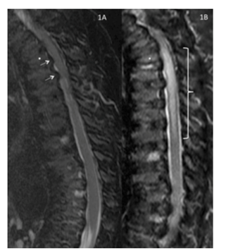
Sagittal T2/short T1 inversion recovery – weighted image of the thoracic and lumbar spine obtained 7 months preoperatively.

Reproduced with permission from Drummond, Can J Anesth, 2015.

Appropriate inter- and postoperative monitoring techniques should consider the operative risk posed by spinal cord compression and spinal joint laxity.2

Summary1:

In this illustrative case, postoperative spinal cord injury was disproportionate to the degree of spinal stenosis. This case is an example of the exaggerated vulnerability of people with Morquio A to spinal cord compression and damage.

Spinal abnormalities affect all persons with Morquio A.1

Anaesthesiologists should consider subclinical spinal stenosis with spinal column joint laxity adding to the risk of compression.2

Case history1

Preoperative evaluation:
  • 16-year-old patient with Morquio A scheduled for bilateral distal femoral osteotomies to relieve genu valgum deformities
  • Due to several episodes of faecal incontinence, patient received neurosurgical examination revealing
    • Normal lower extremity strength and reflexes
    • Slow, ataxic gait consistent with musculoskeletal abnormalities
  • Magnetic resonance imaging (MRI) showed
    • Cervical findings of hypoplastic odontoid process and mild or moderate stenosis from C2 through C6 with ventral contour deformities of the cervical spinal cord
    • Thoracic (T) findings of moderate stenosis from T1-2 to T3-4 with slight kyphosis at the thoracolumbar junction
    • No obvious lumbar (L) findings
    • Minimal cerebrospinal fluid around the cord T11-L1
  • The absence of cord signals suggesting myelomalacia was noted
  • Flexion-extension X-rays showed no atlantoaxial instability
  • Patient experienced moderately severe sleep apnoea requiring continuous positive airway pressure at home
  • Neurosurgical intervention was deemed unnecessary


case_study2.2

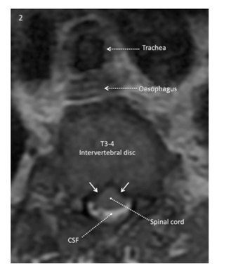
The axial T2-weighted image at the level of the T3-4 intervertebral disk obtained 7 months preoperatively shows cerebrospinal fluid (white) around the posterior portion of the spinal cord but not anterior. The anterior surface of the cord has a “tented” rather than a smooth convex configuration, reflecting bilateral ventrolateral pressure on the anterior surface of the cord (solid white arrow).

Reproduced with permission from Drummond, Can J Anesth, 2015.

Intraoperative care1:
  • Blood pressure (BP) was 110/83 mm Hg at admission
  • Catheter was uneventfully inserted 5 cm into the epidural space through the L2-3 interspace while patient was in a sitting position
  • Patient moved to a supine position
  • General anaesthesia was induced
  • Laryngoscopy allowed for uncomplicated placement of endotracheal tube
  • BP was recorded at 5-minute intervals
  • Transient hypertension was monitored but no vasopressors were administered
Post anaesthesia monitoring1:
  • In the PACU, 5 hours after the case began, absence of lower extremity movement was recorded
  • Epidural infusion was discontinued 30 minutes later
  • At 6.5 hours an examination revealed the absence of motor function and sensation below T4
  • After 9 hours the catheter was removed
  • At 13.5 hours, motor and sensory levels were noted to be at T5
  • Epidural haematoma was suspected and an MRI was ordered
  • MRI was performed 21 hours after arrival in the PACU
    • MRI findings revealed
      • Abnormal T2/short T1 inversion recovery cord signal from T1 through T7
      • Restricted diffusion of full diameter of the spinal cord
  • The patient remained paraplegic at the T4 level

Hypothetical case study: Perceived delayed development, diagnosis and enzyme replacement therapy (ERT) in a patient with mucopolysaccharidosis type VI (MPS VI)1,a

Based on a case study provided by Dr Zakharchuk

Perceived delayed development, a concussion, and decreased endurance and mobility lead to subsequent diagnosis of MPS VI1

    Description1:

  • In 2004, parents consulted local geneticist about delays in their child’s physical development and microanomalies in development
  • The local geneticist performed cytogenetic testing, which revealed no chromosomal abnormality
  • Seven years after initial presentation, following concussion and extremity weakness, patient was referred to an experienced geneticist and subsequently diagnosed with MPS VI and placed on ERT
  • First year on ERT resulted in significant functional improvements and improved respiratory function
Diagnosis of MPS should be considered in patients with hallmark abnormalities; referral to a geneticist centre with experience in testing for MPS can improve time to diagnosis and enable subsequent ERT.2

Case history1

2004:
  • Parents consulted geneticist about developmental delays
  • Cytogenetic examination was conducted
    • Results: karyotype 46HH, 9phgh (normal variant, not a sign for chromosomal abnormality)
2010:
  • Patient experienced a closed traumatic brain injury with concussion
  • Complaints of weakness in the left-side extremities
2011:
  • Decrease in endurance and mobility; patient unable to move independently
  • Enzyme test performed and MPS VI diagnosis confirmed
    • Activity of lysosomal α-L-iduronidase (IDUA) in peripheral blood: IDUA – 70/2 nmol/mg/18 h
    • Lysosomal arylsulfatase B (ARSB) activity: ARSB 9.3 nmol/mg/h
  • Cervical spinal surgery required; С1-С2-С3, Summit Si–construction type was used
2013:
  • Patient began ERT, with decreased glycosaminoglycan (GAG) levels and improvements in functional and respiratory test performance
aHearing loss is common and may lead to a perception that the patient may have developmental delay.3

bIndividual outcomes in patients receiving ERT may vary.
Clinical measure
Baseline measurement
ERT assessment 1 year
12MWT
150 steps
670 m
3MSC
5 steps
80 steps
uGAG
614 μg/mL
311 μg/mL
FVC
Unable to perform
31%
FEV1
Unable to perform
33%
Abbreviations: 3MSC, 3-minute stair climb; 12MWT, 12-minute walk test; FEV1, forced expiratory volume in 1 second; FVC, forced vital capacity; GAG, glycosaminoglycan.

Summary1

  • Delayed diagnosis of MPS leads to lack of intervention and disease-specific management, resulting in poor patient outcomes
  • ERT can improve functional and respiratory measures

Early diagnosis is key to initiating ERT, when available, and providing opportunities to improve patient outcomes.4-7,b As this case demonstrates, ERT has the potential to improve key clinical parameters, such as endurance and respiratory measures, which may be critical to patient quality of life, maintenance of ambulation, and activities of daily living.8,9

Case study: Rapid progression of developmental delay, coupled with parallel symptomatology in younger sibling, leads to dual diagnoses of mucopolysaccharidosis IIIA (MPS IIIA)1

Age at diagnosis: 12 years

Time to diagnosis: 11 years

Key diagnostic symptom: Motor and speech delays

Diagnosing doctor: Geneticist

Onset of neurological symptoms at 1 year of age, followed by rapid progression of developmental delay, leads to diagnosis of MPS IIIA in 2 siblings1

    Description1:

  • Two siblings were investigated clinically and genetically for progressive neurodegenerative disease, developmental and motor delays, and behavioural abnormalities
  • Normal development up to 1 year of age in eldest daughter; progressive motor and speech delays prompted multiple visits to a paediatric neurologist
  • Metabolic screening, including glycosaminoglycans (GAGs), karyotype testing, and MRIs, was normal
  • Similar rapid progression of symptoms in both siblings, including severe spasticity and intellectual disability with autistic features and incontinence, prompted genetic testing and subsequent diagnosis of MPS IIIA at ages 12 and 10
Biochemical testing may be misleading; direct referral to a geneticist and/or metabolic centre is recommended if hallmark clinical features present and progress.1

    Summary1:

  • Rapid progression of developmental delay, coupled with similar signs and symptoms in younger sister, led to eventual referral to a geneticist
  • Metabolic screening tools such as urinary GAGs may not be reliable – in this case, for example, delay in genetic testing resulted in a time to diagnosis of 11 years

As this case demonstrates, signs and symptoms can progress rapidly, particularly with neurological deterioration and worsening developmental delay in patients with MPS IIIA. For families with multiple children demonstrating any features indicative of MPS, the index of suspicion should be especially high.1

While continual monitoring and evaluation are critical, early intervention through direct referral to a geneticist and/or metabolic centre, rather than metabolic screening using urinary GAGs, is recommended to expedite diagnosis.1

Case history1

Age 1:
  • Initial signs and symptoms of motor and speech delays start
  • Referred to a paediatric neurology clinic at 18 months for suspected motor delay. Examination revealed
    • Mild motor delay but could stand with support and speak a few words
    • Hypertonia of the lower extremities and increased tendon reflexes of lower and upper limbs
    • No dysmorphic features and no visceral enlargement; cranial nerves, sensory perception, and cerebellar function were also normal
  • Referred for metabolic screening, including GAGs in the urine (uGAGs), which revealed normal uGAG levels
Age 2:
  • Additional testing carried out, showing normal test results in
    • Karyotype testing and brain magnetic resonance imaging (MRI)
    • Cerebrospinal fluid examination for cell count and protein, glucose and lactate levels
Age 3:
  • Paediatric neurology and child development clinic followed up and performed a second uGAG screening, which was found to be normal
Age 11:
  • Patient started suffering from recurrent convulsions, paroxysmal events of generalised hypertonia, and absences with loss of consciousness for several minutes
  • Electroencephalography record was normal; patient treated with valproic acid with good response
Age 12:
  • Neurological signs rapidly progressed, including severe spasticity, impaired dexterity, and severe intellectual disability with autistic features and incontinence
  • Additional signs and symptoms included hearing impairment, joint contractures, pes cavus, and skin discoloration of the arms, hands, legs and feet
  • A second MRI revealed parieto-occipital atrophy, including cortex atrophy, thin corpus callosum, white matter thinning, and mild ventriculomegaly
  • Similar symptom presentation and progression in sibling prompted genetic testing and subsequent diagnosis of MPS IIIA; uGAG screening also demonstrated increased levels of uGAGs

case_study4.1

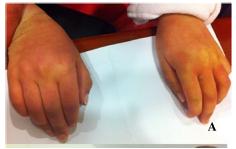
Examination reveals clear skin discolouration in patient with MPS IIIA.1

case_study4.2

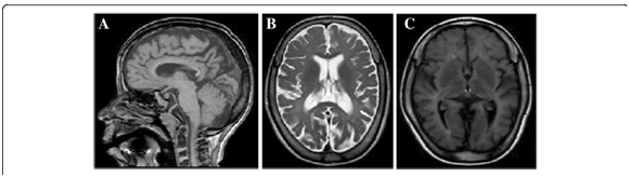
MRI revealed diffuse hypomyelination, thinning of the corpus callosum, and moderate cerebral atrophy, symptoms that developed over time.1

Optimise patient outcomes through coordinated management.

It's a new era in management. Stay informed.

References:  1. Data on file. Biomarin Pharmaceutical.  2. Hendriksz CJ, Berger KI, Giugliani R, et al. International guidelines for the management and treatment of Morquio A syndrome. Am J Med Genet Part A. 2014;9999A:1-15. doi:10.1002/ajmg.a.36833.  3. Giugliani R, Lampe C, Guffon N, et al. Natural history and galsulfase treatment in mucopolysaccharidosis VI (MPS VI, Maroteaux-Lamy syndrome)–10-year follow-up of patients who previously participated in an MPS VI Survey Study. Am J Med Genet A. 2014;164A(8):1953-1964. doi:10.1002/ajmg.a.36584.

References:  1. Drummond JC, Krane EJ, Tomatsu S, Theroux MC, Lee RR. Paraplegia after epidural-general anesthesia in a Morquio patient with moderate thoracic spinal stenosis. Can J Anesth. 2015;62(1):45-49. doi:10.1007/s12630-014-0247-1.  2. Spinello CM, Novello LM, Pitino S, et al. Anesthetic management in mucopolysaccharidoses. ISRN Anesthesiol. 2013;2013:1-10. doi:10.1155/2013/791983.

References:  1. Data on file. Biomarin Pharaceutical.  2. Hendriksz CJ, Berger KI, Giugliani R, et al. International guidelines for the management and treatment of Morquio A syndrome. Am J Med Genet Part A. 2014;9999A:1-15. doi:10.1002/ajmg.a.36833.  3. Valayannopoulos V, Nicely H, Harmatz P, Turbeville S. Mucopolysaccharidosis VI. Orphanet J Rare Dis. 2010;5:5. doi:10.1186/1750-1172-5-5.  4. Clarke LA. Pathogenesis of skeletal and connective tissue involvement in the mucopolysaccharidoses: glycosaminoglycan storage is merely the instigator. Rheumatology (Oxford). 2011;50(suppl 5):v13-18. doi:10.1093/rheumatology/ker395.  5. Lehman TJA, Miller N, Norquist B, Underhill L, Keutzer J. Diagnosis of the mucopolysaccharidoses. Rheumatology. 2011;50(suppl 5):v41-v48.  6. Morishita K, Petty RE. Musculoskeletal manifestations of mucopolysaccharidoses. Rheumatology. 2011;50(suppl 5):v19-v25. doi:10.1093/rheumatology/ker397.  7. Muenzer J. Overview of the mucopolysaccharidoses. Rheumatology. 2011;50:v4-v12. doi:10.1093/rheumatology/ker394.  8. Hendriksz C. Improved diagnostic procedures in attenuated mucopolysaccharidosis. Br J Hosp Med. 2011;72(2):91-95.  9. Muenzer J. Early initiation of enzyme replacement therapy for the mucopolysaccharidoses. Mol Genet Metab. 2014;111(2):63-72. doi:10.1016/j.ymgme.2013.11.015.

Reference:  1. Sharkia R, Mahajnah M, Zalan A, Sourlis C, Bauer P, Schöls L. Sanfilippo type A: new clinical manifestations and neuro-imaging findings in patients from the same family in Israel: a case report. J Med Case Rep. 2014;8:78. doi:10.1186/1752-1947-8-78.