X
X
X
X
Back to Top

“MPS disorders have variable presentation and a wide spectrum of severity. They may present as a single organ dysfunction or a common paediatric problem. The condition can be easily missed even amongst the specialists. Without high index of suspicion and detailed work up, they can be missed for years even after surgery”. – Dr Choy Yew Sing

Recognise the possibility – raise your suspicion

In many genetic conditions, diagnostic delay is common due to lack of clinical suspicion

Many patients with mucopolysaccharidosis (MPS) disorders experience significant diagnostic delay because of disease rarity, phenotypic heterogeneity, and a myriad of seemingly unrelated symptoms associated with the disease.1-3 In a recent survey of patients with rare diseases (N=920) sponsored by the Global Genes Project, the mean reported length of time from symptom onset to accurate diagnosis was 4.8 years, with a range of 0 to 20 years.4 Commonly misperceived as musculoskeletal conditions, MPS disorders are a group of inherited enzyme deficiencies that manifest in progressive, multisystemic, and life-threatening complications.5-7

Watch key opinion leader Dr Offiah talk about variability in phenotype and disease progression.

The emergence of specific therapies for some MPS disorders has increased the need for accurate and early diagnosis.1,6,8-10 Any delay in diagnosis or treatment may result in the following:

  • Serious systemic complications11,12
  • Irreversible organ damage12
  • Delay in initiating enzyme replacement therapy (ERT) to address underlying enzyme deficiency2
  • Lack of access to disease-specific management, with concomitant increase in risk of surgical mortality1,6,9,10,13
Since treatment exists for several MPS disorders, delay in diagnosis can mean delay in therapy and/or delay in disease-specific management.6,9,10

MPS: hiding in plain sight

Early recognition hinges on early suspicion

MPS disorders are clinically heterogeneous and result in serious complications regardless of the rate of progression, which can range from slowly to rapidly progressing. While rates of progression are gross characterisations of disease severity, patients with slowly progressing disease are also at risk for the serious morbidities and mortality commonly associated with rapidly progressing disease.2,6,14

  • If untreated, many individuals do not live far into adulthood (second and third decade).15
  • Individuals with slowly progressing disease typically present with symptoms later in life.14,16

Patients with MPS can present with classical or non-classical patterns of signs and symptoms.16

  • Classical symptomology may present as more easily recognisable patterns of signs and symptoms that have been extensively documented in literature and in clinical practice.16
  • Non-classical symptomology may present more subtly – without the distinct facial features, short stature, or obvious musculoskeletal involvement of classical forms of MPS – making this phenotype more difficult to distinguish from other, more common skeletal or metabolic disorders.16
  • As an example, in an international registry, approximately 25% of patients with Morquio A syndrome.27
  • overview1.1
    Non-classical patient
  • overview1.2
    Non-classical patient

Slowly progressing disease – and/or non-classical phenotypic appearance – can belie severe clinical course in specific organ progression.18 Regardless of phenotype, symptoms can progress into end-organ damage.12

Overt and generally observable signs should raise your suspicion

Regardless of the clinical setting, there may be overt and generally observable signs that should raise your suspicion. Upon further examination, additional symptomatology may be discovered through specialty-specific targeted clinical assessments, laboratory findings and patient history. This division is illustrated below.

Signs and symptoms of MPS1,2,5,11,12,16-28

Musculoskeletal

General features

  • Abnormal gait
  • Bone dysplasia
  • Claw hands
  • Coarse facial features
  • Joint pain
  • Macrocephaly
  • Pectus carinatum
  • Reduced endurance/exercise intolerance
  • Short stature/growth retardationa

Features revealed by specialty–specific assessment

  • Abnormal gait
  • Bone deformities
  • Dysostosis multiplex
  • Genu valgum
  • Joint involvement (contractures, joint laxity) without inflammation
  • Spinal subluxation

Rheumatological

General features

  • Decreased joint mobility
  • Hip stiffness and pain
  • Joint pain
  • Joint stiffness or laxity

Features revealed by specialty–specific assessment

  • Carpal tunnel syndrome
  • Joint involvement without joint swelling or erosive bone lesions

Ear, Nose, and Throat

General features

  • Conductive and/or sensorineural hearing loss
  • Enlarged tongue
  • Otitis media

Features revealed by specialty-specific assessment

  • Abnormal epiglottis
  • Depressed nasal bridge
  • Hypertrophic adenoids
  • Hypertrophic tonsils
  • Middle ear mucus
  • Narrowing of supraglottic and infraglottic airway
  • Ossicular malformation
  • Recurrent and excessive rhinorrhea
  • Recurrent otitis media
  • Tracheal thickening/compression
  • Tubular obstruction
  • Tympanic membrane thickening

Ophthalmological

General features

  • Cataracts
  • Diffuse corneal clouding
  • Glaucoma

Features revealed by specialty-specific assessment

  • Amblyopia
  • Corneal clouding with characteristic “ground glass” appearance
  • High hyperopia
  • Hypertelorism
  • Optic nerve abnormalities (swelling and atrophy)
  • Peripheral vascularisation of the cornea
  • Progressive pseudo-exophthalmos
  • Reduction in visual acuity
  • Retinopathy
  • Strabismus

Neurological

General features

  • Behavioural abnormalities (typically not present in MPS IVA and VI)
  • Developmental delay (typically not present in MPS IVA and VI)
  • Hearing impairment
  • Seizures (typically not present in MPS IVA and VI)

Features revealed by specialty-specific assessment

  • Arachnoid cysts (typically not present in MPS IVA and VI)
  • Brain atrophy (typically not present in MPS IVA and VI)
  • Carpal tunnel syndrome
  • Cervical cord compression/myelopathy/subluxation
  • Enlarged perivascular space
  • Hydrocephalus
  • Odontoid dysplasia
  • Pachymeningitis cervicalis
  • Papilledema/optic atrophy
  • Sensorineural deafness
  • Signal-intensity abnormalities
  • Spinal canal stenosis
  • Ventriculomegaly

Cardiovascular

General features

  • Reduced endurance/exercise intolerance

Features revealed by specialty-specific assessment

  • Pulmonary hypertension
  • Thickened, regurgitant or stenotic mitral or aortic valves in presence of left ventricular hypertrophy
  • Tricuspid regurgitation

Pulmonary

General features

  • Reduced endurance/exercise intolerance
  • Sleep apnoea

Features revealed by specialty-specific assessment

  • Obstructed upper and lower airways (bronchial narrowing, narrowing of supraglottic and infraglottic airway)
  • Progressive reduction in lung volume
  • Respiratory infections
  • Sleep disorders (obstructive sleep apnoea/hypopnoea syndrome and upper airway resistance syndrome)

Gastrointestinal

General features

  • Abdominal pain
  • Constipation
  • Hepatosplenomegaly
  • Hernias
  • Loose stools

Features revealed by specialty-specific assessment

  • Hepatosplenomegaly

Dental

General features

  • Abnormal buccal surfaces
  • Dentinogenesis imperfecta
  • Hypodontia
  • Pointed cusps
  • Spade–shaped incisors
  • Thin enamel

Features revealed by specialty-specific assessment

  • Abnormal buccal surfaces
  • Thin enamel

aSkeletal involvement and short stature may be less overt in some patients.

As skeletal manifestations are common among individuals with MPS, these disorders may resemble or present as other musculoskeletal conditions.1,10,16 MPS should be considered in a variety of clinical settings, including the following16,29:

  • Skeletal dysplasia clinics
  • Orthopaedics
  • Paediatric rheumatology
  • Metabolic bone clinics
Cognitive and mental impairment are not hallmark signs of all MPS disorders. Never rule out MPS based on the absence of cognitive/mental impairment.1

Prompt referral and diagnosis is the best chance to improve patient lives1,6; understand the key clinical hallmarks of MPS and refer suspected patients immediately to a geneticist or metabolic centre for diagnostic testing.1,16,29

Patients with MPS are diverse – discover the full spectrum

In your everyday practice, diverse presentations can complicate patient diagnosis

The best way to accurately diagnose MPS is to refer a patient to a geneticist who is familiar with MPS and knows how to confirm the diagnosis.1,19

You should suspect MPS when confronted by patients who

  • Exhibit any number or pattern of heterogeneous and seemingly unrelated signs1,19
  • Have high surgical need and burden1,19

When you suspect MPS, refer immediately to a geneticist or metabolic centre.1,19

Early identification relies on seeing the full clinical picture and knowing the key clinical hallmarks of MPS.1,2,6,10,19

Discover the pathology behind the presentation

MPS is a category of lysosomal storage disorders. There are 11 identified MPS disorders with varying average ages of onset, which may not correlate to age of diagnosis. Each one is caused by a deficiency of a lysosomal enzyme.1,2,6,10,19

MPS disorders exhibit similar pathologies and mechanisms, regardless of the specific enzyme deficiency.1,6

  • Lysosomal enzymes break down glycosaminoglycans (GAGs).1,6
  • Accumulation of GAGs throughout the body causes progressive damage manifesting in a range of multisystemic consequences.1,6

As an example, the video below illustrates the mechanism of disease for Morquio A.

Optimise outcomes – change the clinical course for your patients

A coordinated, multidisciplinary care-delivery model

Decades of disease and therapeutic research and practice have culminated in new approaches to clinical management of MPS disorders. At the centre of today’s new era of management is a coordinated, multidisciplinary care-delivery model-centred around the geneticist or metabolic specialist.

The unique risks and needs associated with these multisystemic, complex conditions are best addressed by coordinating three pillars of care: enzyme replacement therapy (ERT) (if available), procedural care, and lifetime management.2,6,8,36-38

As the management paradigm continues to evolve, changes in best practices and strategies continue to show greater promise for the patients and families affected by the disorders.8,33

Prompt referral for diagnosis is essential to optimise care for patients with MPS.1,2,6

Read More

Early investigation leads to early intervention.
Avoid delay.

It's a new era in management. Stay informed.

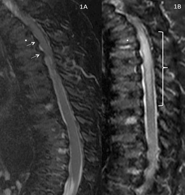
References:  1. Lehman TJA, Miller N, Norquist B, Underhill L, Keutzer J. Diagnosis of the mucopolysaccharidoses. Rheumatology. 2011;50(suppl 5):v41-v48.  2. Hendriksz C. Improved diagnostic procedures in attenuated mucopolysaccharidosis. Br J Hosp Med. 2011;72(2):91-95.  3. Hendriksz CJ, Al-Jawad M, Berger KI, et al. Clinical overview and treatment options for non-skeletal manifestations of mucopolysaccharidosis type IVA. J Inherit Metab Dis. 2013;36(2):309-322. doi:10.1007/s10545-012-9459-0.  4. Engel PA, Bagal S, Broback M, Boice N. Physician and patient perceptions regarding physician training in rare diseases: the need for stronger educational initiatives for physicians. J Rare Disord. 2013;1(2):1-15.  5. Clarke LA, Winchester B, Giugliani R, Tylki-Szymańska A, Amartino H. Biomarkers for the mucopolysaccharidoses: discovery and clinical utility. Mol Genet Metab. 2012;106(4):396-402. doi:10.1016/j.ymgme.2012.05.003.  6. Muenzer J, Beck M, Eng CM, et al. Long-term, open-labeled extension study of idursulfase in the treatment of Hunter syndrome. Genet Med. 2011;13(2):95-101. doi:10.1097/GIM.0b013e3181fea459.  7. Muenzer J. Early initiation of enzyme replacement therapy for the mucopolysaccharidoses. Mol Genet Metab. 2014;111(2):63-72. doi:10.1016/j.ymgme.2013.11.015.  8. Muenzer J, Wraith JE, Clarke LA, International Consensus Panel on the Management and Treatment of Mucopolysaccharidosis I. Mucopolysaccharidosis I: management and treatment guidelines. Pediatrics. 2009;123(1):19-29. doi:10.1542/peds.2008-0416.  9. Clarke LA. Pathogenesis of skeletal and connective tissue involvement in the mucopolysaccharidoses: glycosaminoglycan storage is merely the instigator. Rheumatology (Oxford). 2011;50(suppl 5):v13-18. doi:10.1093/rheumatology/ker395.  10. Morishita K, Petty RE. Musculoskeletal manifestations of mucopolysaccharidoses. Rheumatology. 2011;50(suppl 5):v19-v25. doi:10.1093/rheumatology/ker397.  11. Hendriksz CJ, Berger KI, Giugliani R, et al. International guidelines for the management and treatment of Morquio A syndrome. Am J Med Genet Part A. 2014;9999A:1-15. doi:10.1002/ajmg.a.36833.  12. Berger KI, Fagondes SC, Giugliani R, et al. Respiratory and sleep disorders in mucopolysaccharidosis. J Inherit Metab Dis. 2013;36(2):201-210. doi:10.1007/s10545-012-9555-1.  13. Spinello CM, Novello LM, Pitino S, et al. Anesthetic management in mucopolysaccharidoses. ISRN Anesthesiol. 2013;2013:1-10. doi:10.1155/2013/791983.  14. Lampe C. Attenuated mucopolysaccharidosis: are you missing this debilitating condition? Rheumatology (Oxford). 2012;51(3):401-402. doi:10.1093/rheumatology/ker375.  15. Tomatsu S, Montaño AM, Oikawa H, et al. Mucopolysaccharidosis type IVA (Morquio A disease): clinical review and current treatment: a special review. Curr Pharm Biotechnol. 2011;12(6):931-945. doi:1389-2010/11.  16. Lachman RS, Burton BK, Clarke LA, et al. Mucopolysaccharidosis IVA (Morquio A syndrome) and VI (Maroteaux-Lamy syndrome): under-recognized and challenging to diagnose. Skeletal Radiol. 2014;43(3):359-369. doi:10.1007/s00256-013-1797-y.  17. Montaño AM, Tomatsu S, Gottesman GS, Smith M, Orii T. International Morquio A Registry: clinical manifestation and natural course of Morquio A disease. J Inherit Metab Dis. 2007;30(2):165-174. doi:10.1007/s10545-007-0529-7.  18. Thümler A, Miebach E, Lampe C, et al. Clinical characteristics of adults with slowly progressing mucopolysaccharidosis VI: a case series. J Inherit Metab Dis. 2012;35(6):1071-1079. doi:10.1007/s10545-012-9474-1.  19. Muenzer J. The mucopolysaccharidoses: a heterogeneous group of disorders with variable pediatric presentations. J Pediatr. 2004;144(suppl 5):S27-S34.  20. Kinirons MJ, Nelson J. Dental findings in mucopolysaccharidosis type IV A (Morquio’s disease type A). Oral Surg Oral Med Oral Pathol. 1990;70(2):176-179.  21. Lachman R, Martin KW, Castro S, Basto MA, Adams A, Teles EL. Radiologic and neuroradiologic findings in the mucopolysaccharidoses. J Pediatr Rehabil Med. 2010;3(2):109-118. doi:10.3233/PRM-2010-0115.  22. Cimaz R, Coppa GV, Koné-Paut I, et al. Joint contractures in the absence of inflammation may indicate mucopolysaccharidosis [hypothesis]. Pediatr Rheumatol Online J. 2009;7:18. doi:10.1186/1546-0096-7-18.  23. Fahnehjelm KT, Ashworth JL, Pitz S, et al. Clinical guidelines for diagnosing and managing ocular manifestations in children with mucopolysaccharidosis. Acta Ophthalmol. 2012;90(7):595-602. doi:10.1111/j.1755-3768.2011.02280.x.  24. Zafeiriou DI, Batzios SP. Brain and spinal MR imaging findings in mucopolysaccharidoses: a review. AJNR Am J Neuroradiol. 2013;34(1):5-13. doi:10.3174/ajnr.A2832.  25. Braunlin EA, Harmatz PR, Scarpa M, et al. Cardiac disease in patients with mucopolysaccharidosis: presentation, diagnosis and management. J Inherit Metab Dis. 2011;34(6):1183-1197. doi:10.1007/s10545-011-9359-8.  26. Braunlin E, Orchard PJ, Whitley CB, Schroeder L, Reed RC, Manivel JC. Unexpected coronary artery findings in mucopolysaccharidosis. Report of four cases and literature review. Cardiovasc Pathol. 2014;23(3):145-151. doi:10.1016/j.carpath.2014.01.001.  27. Mesolella M, Cimmino M, Cantone E, et al. Management of otolaryngological manifestations in mucopolysaccharidoses: our experience. Acta Otorhinolaryngol Ital. 2013;33(4):267-272.  28. Martins AM, Dualibi AP, Norato D, et al. Guidelines for the management of mucopolysaccharidosis type I. J Pediatr. 2009;155(4)(suppl 2):S32-S46. doi:10.1016/j.jpeds.2009.07.005.  29. Wood TC, Harvey K, Beck M, et al. Diagnosing mucopolysaccharidosis IVA. J Inherit Metab Dis. 2013;36(2):293-307. doi:10.1007/s10545-013-9587-1.  30. Data on file. Biomarin Pharmaceutical, Inc.  31. Drummond JC, Krane EJ, Tomatsu S, Theroux MC, Lee RR. Paraplegia after epidural-general anesthesia in a Morquio patient with moderate thoracic spinal stenosis. Can J Anesth. 2015;62(1):45-49. doi:10.1007/s12630-014-0247-1.  32. Sharkia R, Mahajnah M, Zalan A, Sourlis C, Bauer P, Schöls L. Sanfilippo type A: new clinical manifestations and neuro-imaging findings in patients from the same family in Israel: a case report. J Med Case Rep. 2014;8:78. doi:10.1186/1752-1947-8-78.  33. Heese BA. Current strategies in the management of lysosomal storage diseases. Semin Pediatr Neurol. 2008;15(3):119-126. doi:10.1016/j.spen.2008.05.005.  34. Kakkis ED. Enzyme replacement therapy for the mucopolysaccharide storage disorders. Expert Opin Investig Drugs. 2002;11(5):675-685.  35. Agency for Healthcare Research and Quality. Defining the PCMH. https://pcmh.ahrq.gov/page/defining-pcmh. Accessed December 15, 2015.  36. Hwu W-L, Okuyama T, But WM, et al. Current diagnosis and management of mucopolysaccharidosis VI in the Asia-Pacific region. Mol Genet Metab. 2012;107(1-2):136-144. doi:10.1016/j.ymgme.2012.07.019.  37. Klitzner TS, Rabbitt LA, Chang RKR. Benefits of care coordination for children with complex disease: a pilot medical home project in a resident teaching clinic. J Pediatr. 2010;156(6):1006-1010. doi:10.1016/j.jpeds.2009.12.012.  38. Mosquera RA, Avritscher EBC, Samuels CL, et al. Effect of an enhanced medical home on serious illness and cost of care among high-risk children with chronic illness: a randomized clinical trial. JAMA. 2014;312(24):2640-2648. doi:10.1001/jama.2014.16419.